| Table of Contents | |
|
Case Report
| ||||||
| Recurrent intestinal obstruction due to Meckel's diverticulum: A case report | ||||||
| Mahir Gachabayov1, Elbrus Abdullaev2, Valentin Babyshin3, Abakar Abdullaev4 | ||||||
|
1MD, Attending Surgeon, Department of Abdominal Surgery, Vladimir City Clinical Hospital of Emergency Medicine, Vladimir, Vladimirskaya Oblast, Russia.
2PhD, Chief Surgeon of Department, Department of Abdominal Surgery, Vladimir City Clinical Hospital of Emergency Medicine, Vladimir, Vladimirskaya Oblast, Russia. 3PhD, Chief Surgeon of Hospital, Vladimir City Clinical Hospital of Emergency Medicine, Vladimir, Vladimirskaya Oblast, Russia. 4MD, Attending Surgeon, Department of Abdominal Surgery, Vladimir City Clinical Hospital of Emergency Medicine, Vladimir, Vladimirskaya Oblast, Russia. | ||||||
| ||||||
|
[HTML Abstract]
[PDF Full Text]
[Print This Article]
[Similar article in Pumed] [Similar article in Google Scholar] |
| How to cite this article: |
| Gachabayov M, Abdullaev E, Babyshin V, Abdullaev A. Recurrent intestinal obstruction due to Meckel's diverticulum: A case report. Edorium J Gastrointest Surg 2015;2:1–5. |
|
Abstract
|
|
Introduction:
Meckel's diverticulum is the most common congenital anomaly of gastrointestinal tract. Complications of Meckel's diverticulum include gastrointestinal bleeding, bowel obstruction and acute diverticulitis.
Case Report: A 34-year-old male patient was admitted to surgical department with intestinal obstruction 4th times during the last three years. Previous episodes of intestinal obstruction were treated conservatively with success. After last admission conservative treatment attempts were unsuccessful, so the patient underwent surgery. On laparotomy Meckel's diverticulum 40 cm proximal to ileocecal valve was found. The reason of intestinal obstruction appeared to be the mesentery of Meckel's diverticulum (mesodiverticulum) compressing adjacent small bowel and leading to intestinal obstruction. Conclusion: Young patients with recurrent small bowel obstruction should be examined for Meckel's diverticulum and if found the patient should be treated surgically. | |
|
Keywords:
Diverticulectomy, Meckel's diverticulum, Mesodiverticulum, Small bowel obstruction, True diverticulum
| |
|
Introduction
| ||||||
|
Being a remnant of not completely involuted vitello enteric duct, Meckel's diverticulum (MD) is a true diverticulum and the most common congenital anomaly of gastrointestinal tract. Its incidence is 1–3% of population with almost equal distribution between males and females. In most cases Meckel's diverticulum is asymptomatic; however, in 16% of patients different complications can occur. They include emergency surgical complications like gastrointestinal bleeding, bowel obstruction, acute diverticulitis, perforation, etc. Intestinal obstruction can develop in different ways, in some cases it resolves spontaneously and can recur. | ||||||
|
Case Report
| ||||||
|
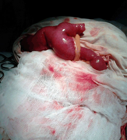
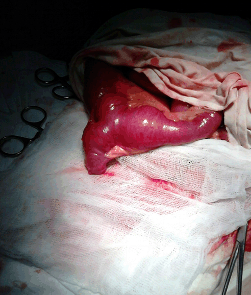
A 34-year-old white male was admitted to Vladimir City Clinical Hospital of Emergency Medicine with clinical manifestation of small bowel obstruction. Abdominal pain, distention, nausea and vomiting started about 10 hours before admission. His past medical history was significant for previous three hospitalizations due to small bowel obstruction. In his discharge cards the diagnosis was shown as Ogilvie syndrome with adynamic ileus. three previous hospitalizations were not long (the longest being three days) and the patient had treated conservatively with success. He had no previous abdominal surgery. On admission, slight distention of abdomen, tenderness in lower quadrants and succussion splash was noted. Abdominal X-ray showed small bowel air-fluid levels and dilatation of small bowel up to 3 cm. On ultrasonography, signs of intestinal obstruction were found. Conservative treatment including body fluid replacement and enemas started immediately. After 10 hours of conservative treatment success was not achieved. The patient underwent surgery. On midline laparotomy mesodiverticulum compressing adjacent ileum and leading to small bowel obstruction appeared to be the reason of intestinal obstruction (Figure 1) (Figure 2) (Figure 3). Meckel's diverticulum with the length of 5 cm was located 40 cm proximal to the ileocecal valve. Its walls were smooth and homogenous on palpation. Diverticulectomy was performed. On pathohistology no ectopic tissue was found. Postoperative period was without any complications. The patient was discharged on eighth postoperative day. | ||||||
| ||||||
| ||||||
| ||||||
|
Discussion
| ||||||
|
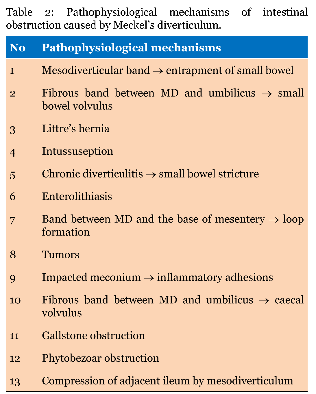
History: Meckel's diverticulum (MD) was first correctly described and interpreted to be the intestinal remnant of the ductus umbilical-intestinalis in 1809 by German anatomist Johann Friedrich Meckel the Younger (1781–1833) [1]. Anatomy: Meckel's diverticulum is located on the antimesenteric wall of ileum 40–60 cm proximal to ileocecal valve. Its average length is 3–5 cm. However, there are reports of mesenteric location [2] and longer MD up to 100 cm. Walls of MD has all 3 layers as small bowel wall. It can contain ectopic gastric or pancreatic tissue in 50% and 5%, respectively. In 5% of cases duodenal or jejunal mucosa can exist. Commonly its orifice is wide. Meckel's diverticulum has its own mesentery and blood supply originating from the branches of superior mesenteric artery. Meckel's diverticulum is commonly located in right lower quadrant of abdominal cavity. That's why its inflammation mimics acute appendicitis. In some cases MD has a mesodiverticular band (a fibrous tubular structure combining mesodiverticulum to parietal peritoneum) which can lead to bowel obstruction and strangulation [3]. Pathology and clinical presentation. Pathology of MD directly correlates with its clinical manifestations. The classification of MD with its complications is given in Table 1. Asymptomatic MD is found eventually on radiologic imaging or intraoperatively during surgery for other abdominal condition. Complications of asymptomatic MD can be ulcers, tumors, umbilical cysts or sinuses without pain and discharge, enteroliths without bowel obstruction and fistulas like vesico-diverticular fistula [4]. Patients with non-complicated symptomatic MD have recurrent abdominal pain. Complications of symptomatic MD are given in Table 1. Among them three complications are surgical emergencies: bleeding, intestinal obstruction and diverticulitis (with or without perforation, abscess and peritonitis). Bleeding is the most common manifestation of MD in children with the highest incidence in two-year-old children. However, bleeding of MD origin can also be encountered in older children and adults. Pathogenesis is alteration of mucosa in and around MD by acidic or alkaline secretion from ectopic gastric or pancreatic tissues. Besides this, chronic inflammation of diverticular mucosa is another reason of bleeding (mostly minor). Chronic minor bleeding from MD can lead to iron deficiency anemia. The second most common in children and the most common in adults complication of symptomatic MD is intestinal obstruction. Pathophysiological mechanisms of intestinal obstruction due to MD are given in Table 2 [5]. In our patient, the pathophysiological mechanism of intestinal obstruction was mesodiverticulum compressing adjacent ileum. Mesodiverticulum compressing adjacent ileum leads to partial bowel obstruction, so mostly can be treated conservatively and can recur several times. The third most common complication of MD is its inflammation- acute diverticulitis. Clinical presentation of acute diverticulitis is similar to that of acute appendicitis because of its location, similar anatomic structure and similar complications. The complications of acute diverticulitis are perforation, local and generalized peritonitis. Pathogenesis of acute diverticulitis include several mechanisms (lumen obstruction, peptic or pressure ulceration, mesenteric edema, torsion, arterial thrombosis) leading to the inflammation of bowel wall starting from mucous layer towards surrounding layers caused by bacterial translocation. Examination. Laboratory and instrumental examination strongly depends on dominating clinical syndrome. For example, for gastrointestinal bleeding syndrome and intestinal obstruction syndrome initial survey and primary examination will be completely different. Endoscopic procedures are of less accuracy for visualization of MD. However, they are helpful to rule out other etiologies of gastrointestinal bleeding. There are reports clearly demonstrating and illustrating MD by wireless capsule endoscopy [6]. However, capsule endoscopy is more accurate and informative for "silent" MD (not in emergency cases). Radiologic imaging is more important for the diagnosis of MD. Technetium-99m scan is an accurate radiologic test based on detection of ectopic gastric mucosa in children with sensitivity of 85% and even higher specificity of 95% [7]. This test is not used in adults because its specificity is extremely low (not more than 9%) [8]. There are three conditions when Meckel's diverticulum can be diagnosed and becomes a subject for surgery: asymptomatic occasionally found during surgery, symptomatic non-complicated (chronic abdominal pain) and symptomatic complicated MD. Among them only symptomatic non-complicated MD can be examined by technetium-99m scan. Among surgical complications of MD acute diverticulitis is the condition which can be diagnosed by ultrasonography and computed tomography (CT) scan. Meckel's diverticulum complicated with intestinal obstruction can be visualized in some cases only by CT scan. Besides this, in some cases CT scan can determine the site of contrast extravasation in MD bleeding. Among invasive diagnostic procedures angiography is of less importance. So that, it can be helpful only for determining of bleeding site in MD bleeding but its transformation to therapeutic procedure can lead to gangrene of MD [9]. Diagnostic laparoscopy is the most accurate diagnostic procedure with sensitivity and specificity reaching to 100%. However, its invasiveness and the need for general anesthesia make it the last facility in the diagnosis of MD. Treatment. Meckel's diverticulum can be treated only surgically. The surgical procedure is either diverticulectomy or small bowel resection depending on the existence of any inflammatory, ischemic or tumorous changes in adjacent ileum [5]. Surgical approach can also be different depending on complications of MD or existence of facilities and experienced surgeon: laparoscopic, laparoscopy-assisted or laparotomy. | ||||||
| ||||||
| ||||||
|
| ||||||
|
Conclusion
| ||||||
|
Meckel's diverticulum (MD) is a rare cause of intestinal obstruction. Among all possible pathological causes compression of adjacent ileum by MD is even rarer. In young patients with recurrent small bowel obstruction MD should be considered to be one of the reasons. If MD is diagnosed the patient should be treated surgically. | ||||||
|
References
| ||||||
| ||||||
|
[HTML Abstract]
[PDF Full Text]
|
|
Author Contributions
Mahir Gachabayov – Substantial contributions to conception and design, Acquisition of data, Analysis and interpretation of data, Drafting the article, Critical revision of the article, Final approval of the version to be published Elbrus Abdullaev – Analysis and interpretation of data, Revising it critically for important intellectual content, Final approval of the version to be published Valentin Babyshin – Analysis and interpretation of data, Revising it critically for important intellectual content, Final approval of the version to be published Abakar Abdullaev – Acquisition of data, Drafting the article, Revising it critically for important intellectual content, Final approval of the version to be published |
|
Guarantor of submission
The corresponding author is the guarantor of submission. |
|
Source of support
None |
|
Conflict of interest
Authors declare no conflict of interest. |
|
Copyright
© 2015 Mahir Gachabayov et al. This article is distributed under the terms of Creative Commons Attribution License which permits unrestricted use, distribution and reproduction in any medium provided the original author(s) and original publisher are properly credited. Please see the copyright policy on the journal website for more information. |
|
|